Liver Anatomy Concise Medical Knowledge
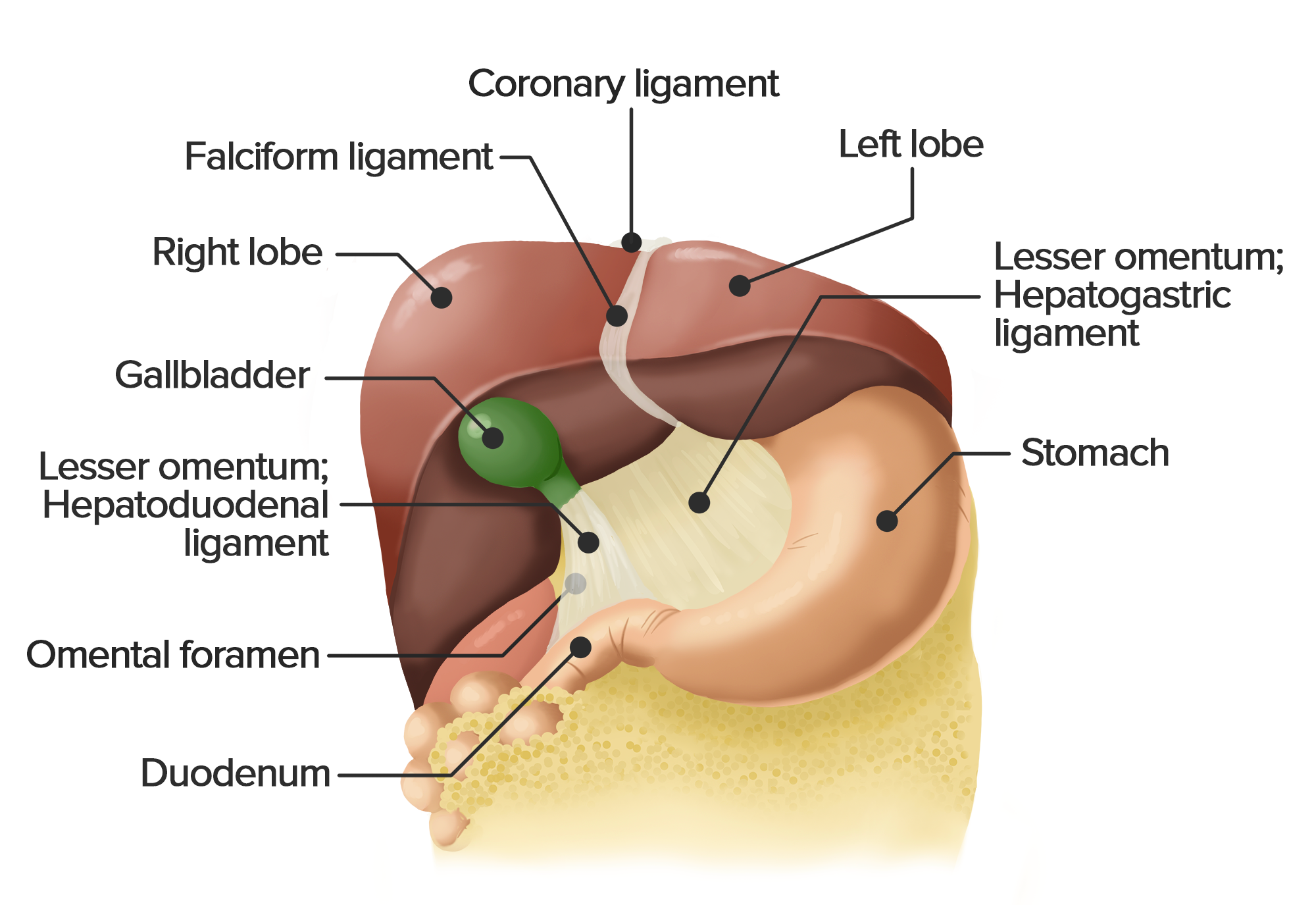
Perihepatic Organs/Anatomy. The gastrointestinal tract has several associations with the liver (illustrated in Fig. 3).The stomach is related to the left hepatic lobe by way of the gastrohepatic ligament or superior aspect of the lesser omentum, which is an attachment of connective tissue between the lesser curvature of the stomach and the left hepatic lobe at the ligamentum venosum.
Liver and gallbladder Anatomy, location and functions Kenhub
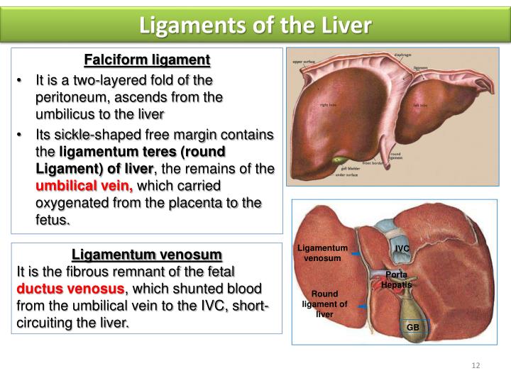
The ligamentum teres (round ligament) represents the obliterated left umbilical vein. It originates from the umbilicus, courses up the anterior abdominal wall within the the peritoneum and then enters the falciform ligament coursing toward the left portal vein and ligamentum venosum.
Liver Anatomy Concise Medical Knowledge
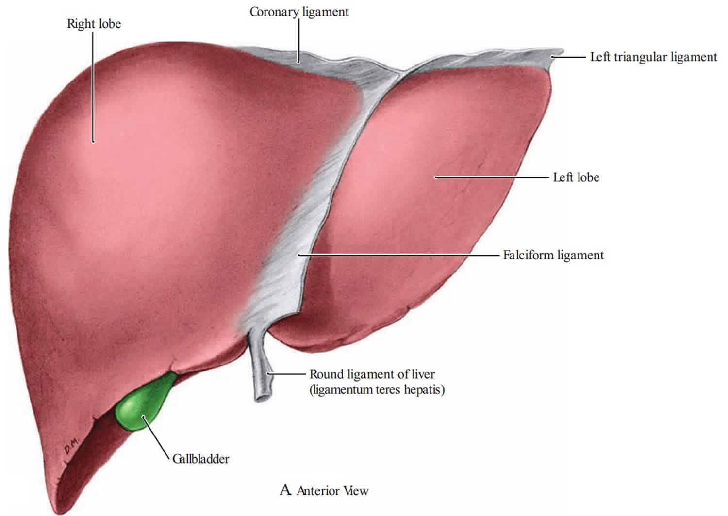
The round ligament of the liver is a ligament that forms part of the free edge of the falciform ligament of the liver. It connects the liver to the umbilicus. It is the remnant of the left umbilical vein. The round ligament divides the left part of the liver into medial and lateral sections.
Ligaments of the gastrointestinal tract Anatomy Kenhub
The liver is a peritoneal organ positioned in the right upper quadrant of the abdomen. It is the largest visceral structure in the abdominal cavity, and the largest gland in the human body. An accessory digestion gland, the liver performs a wide range of functions, such as synthesis of bile, glycogen storage and clotting factor production.. In this article, we shall look at the anatomy of the.
PPT Liver PowerPoint Presentation, free download ID2161297
The round ligament of the liver (or ligamentum teres, or ligamentum teres hepatis) is a ligament that forms part of the free edge of the falciform ligament of the liver. It connects the liver to the umbilicus. It is the remnant of the left umbilical vein. The round ligament divides the left part of the liver into medial and lateral sections.
PPT Liver & Spleen PowerPoint Presentation ID2118838
The two hemilivers are divided on the anterior surface of the liver by the falciform ligament and on the inferior surface by the round ligament as it runs into the umbilical fissure. At the upper margin, the two layers of the falciform ligament divide from each other. On the right side, the falciform ligament attaches the right diaphragmatic.

Surfaces and Bed of Liver Anatomy Human liver anatomy, Liver anatomy, Human anatomy
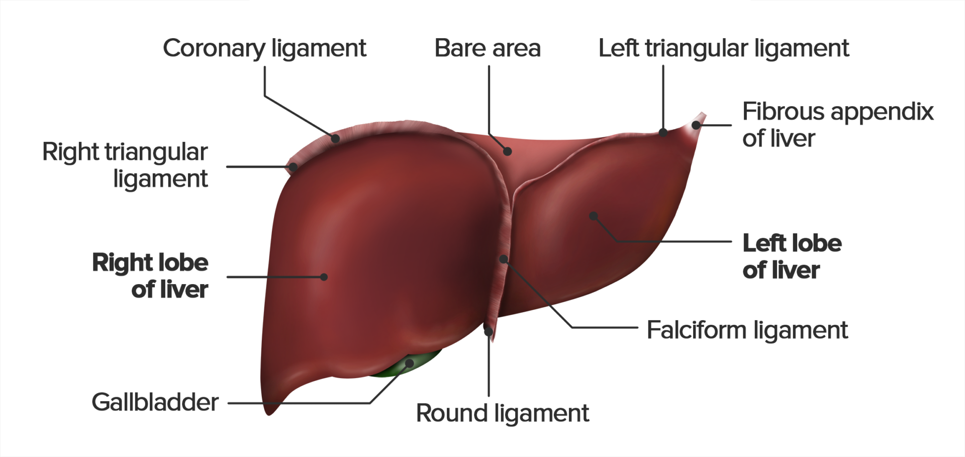
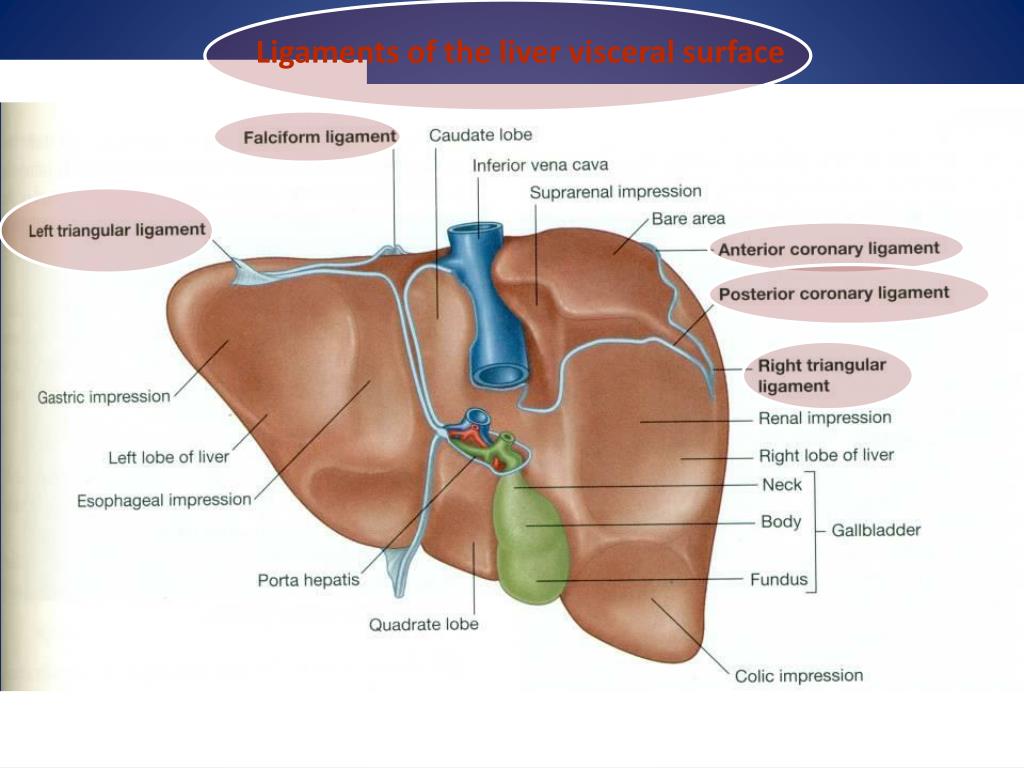
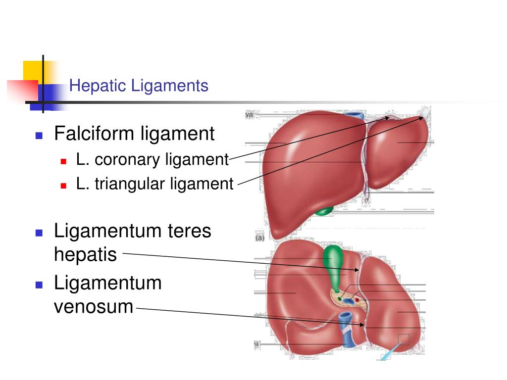
Hepatic ligaments. Several peritoneal ligaments support the position of the liver: round ligament of liver (ligamentum teres), falciform ligament, coronary ligament, triangular ligaments and lesser omentum. The lesser omentum comprises the hepatogastric and hepatoduodenal ligaments which connect the liver to the lesser curvature of the stomach and duodenum.

Gastrointestinal Ligaments USMLE Strike
The free edge of the falciform ligament contains the ligamentum teres hepatis (round ligament of the liver): the obliterated umbilical vein, which is attached to the inferior surface of the liver between segment IV on the right and segment III on the left. The ligamentum venosum (the obliterated ductus venosus) is attached to the inferior.

Inferior aspect of the liver the round ligament continues into the... Download Scientific Diagram
Hepatic portal vein Branches of hepatic portal vein Right branch of hepatic portal vein Left branch of hepatic portal vein Transverse part of left branch of hepatic portal vein Umbilical part of left branch of hepatic portal vein Ligamentum venosum Lateral left branches of hepatic portal vein Umbilical vein Round ligament of liver
Falciform ligament of liver & falciform ligament function
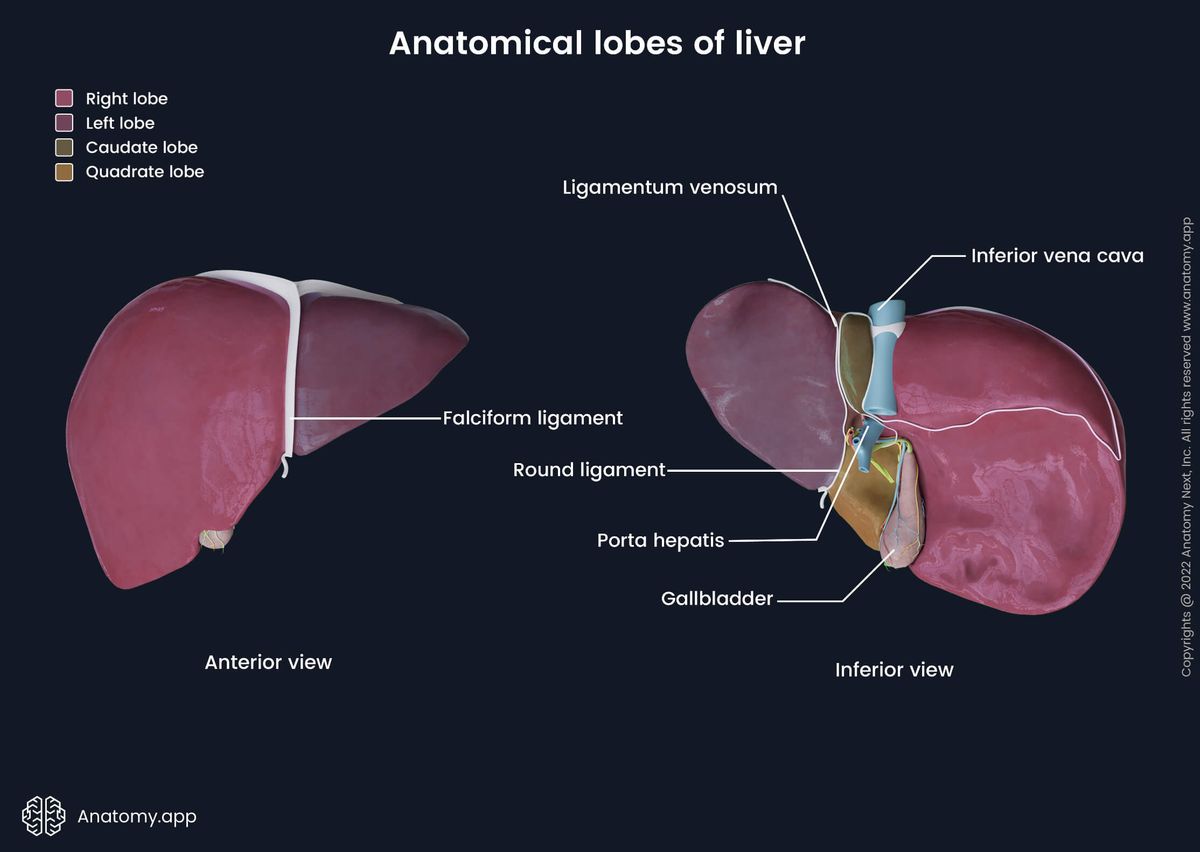
The free-form edge of the falciform ligament contains the round ligament of the liver which is the remnant of the embryonic umbilical vein. Anatomically the liver has four lobes: right, left, caudate, and quadrate.
Ligamentum Teres Hepatis
. Liver parenchyma consists of hepatocytes and hepatic sinusoids . Hepatic sinusoids drain into the central vein of each lobule. The liver is responsible for energy metabolism, synthesis of various substances (e.g., glucose,
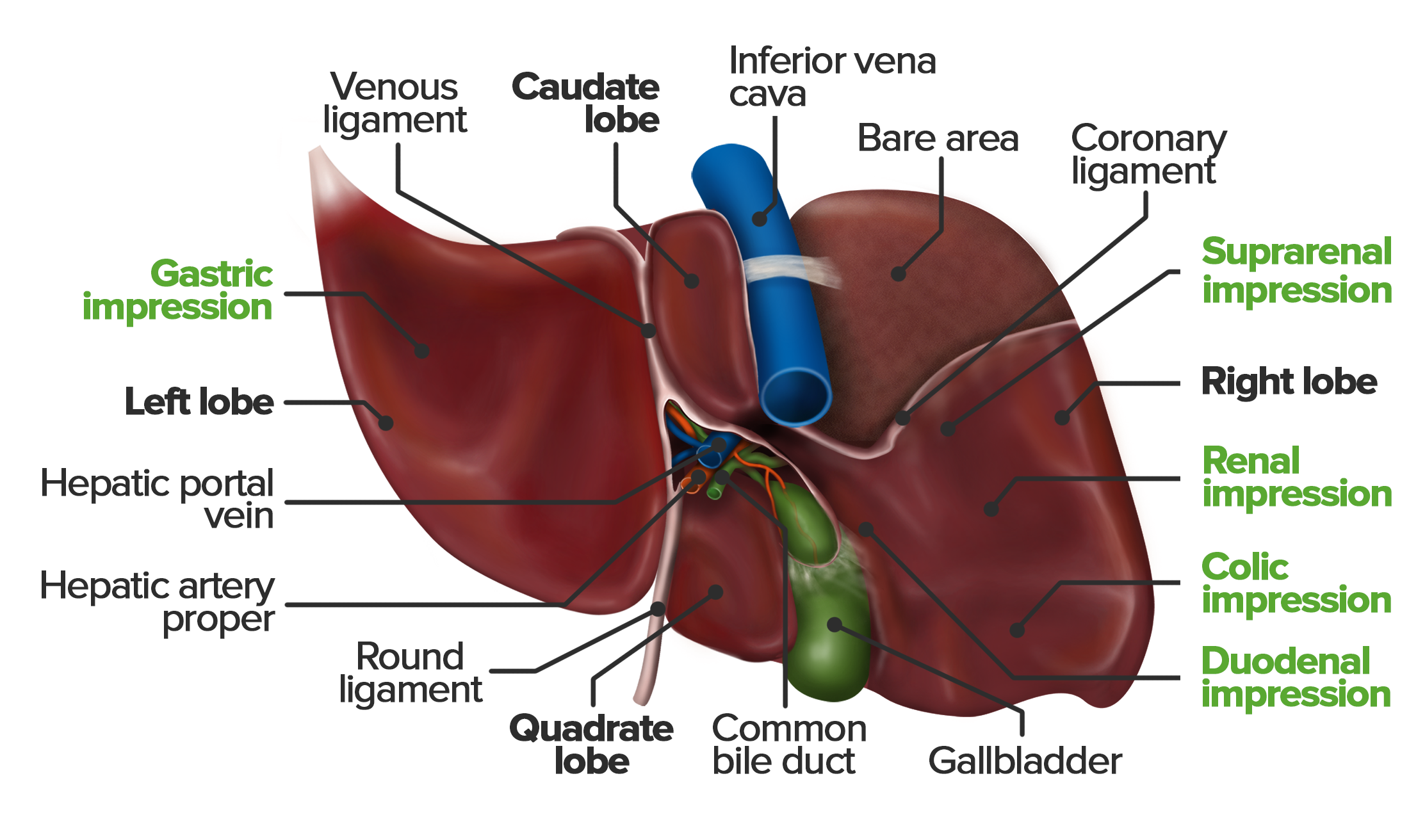
Diagram Of Liver The Liver And Its Functions Center For Liver Disease Transplantation Columbia
12.1.1 Anatomy The hepatic artery, portal vein, and bile ducts (the portal triad) in the ligamentum hepatoduodenale are encased in a membrane and branch, and they constitute Glisson's system. This system consists of extrahepatic and intrahepatic portions.
Liver Encyclopedia Anatomy.app Learn anatomy 3D models, articles, and quizzes
This ligament attaches the liver to the anterior abdominal wall.. The round ligament passes into the groove between the quadrate and left lobe. Remember, above the liver, you've got the diaphragm. I'm drawing the diaphragm on here. This is the diaphragm in red. Reflecting off the diaphragm, you've got folds of peritoneum.
PPT THE LIVER PowerPoint Presentation, free download ID5191228
The round ligament of the liver is the fetal remnant of the umbilical vein, which once traveled from the placenta to the fetal liver to deliver oxygenated blood. [1] Go to: Embryology

Liver Anatomy (Function, Topography, External Structures, Ligaments) YouTube
The bridges comprise the ligaments of the liver as follows: the falciform ligament, right and left coronary ligaments, lesser omentum including the hepatogastric ligament and hepatoduodenal ligament.. The obstructed UV is typically exhibited as a small round-shaped structure that arises from the bifurcation (the umbilical portion) of P3 and.
Liver anatomy (anterior and bottom views) showing the location of... Download Scientific Diagram
Liver ligaments are double-layered folds of peritoneum that attach the liver to surrounding organs, or to the abdominal wall. The majority of ligaments associated with the liver are remnants of embryological blood vessels that regressed as the fetus developed.
